Superionic Ice

What’s an Earth-bound scientist to do when she wants to study superionic ice like the kind found on frozen planets?  Fire up the lasers and make the next best thing herself.

Dr. Federica Coppari, a physicist at Lawrence Livermore National Laboratory, and her co-lead author Marius Millot, used giant lasers to flash freeze water, creating replica superionic ice and snapping images for study..

Federica Coppari with an x-ray diffraction image plate that she and her colleagues used to discover ice XVIII, also known as superionic ice.
Credit:  Eugene Kowaluk/Laboratory for Laser Energetics

The team simply smashed water with laser blasts between diamond anvils.  Using the OMEGA Laser at the University of Rochester – one of the most powerful lasers in the world – they heated the water to around 4,700 degrees Celsius and compress it between 1 and 4 million times the Earth’s atmospheric pressure.  

The 60-beam Omega laser at UR’s Laboratory for Laser Energetics.

Billionths of a second later, as shock waves rippled through and the water began crystallizing into nanometer-size ice cubes, the scientists used 16 more laser beams to vaporize a thin sliver of iron next to the sample. The resulting hot plasma flooded the crystallizing water with X-rays, which then diffracted from the ice crystals, allowing the team to discern their structure.

The atoms in the water had rearranged into the long-predicted but never-before-seen architecture, Ice XVIII: a cubic lattice with oxygen atoms at every corner and the center of each face.  The hydrogen ions float freely within the oxygen lattice. “It’s quite a breakthrough,” Coppari said.

Molecular model of superionic ice XVIII

Unlike the familiar ice found in your freezer or at the north pole, superionic ice is black and hot. A cube of it would weigh four times as much as a normal one. It was first theoretically predicted more than 30 years ago, and although it has never been seen until now, scientists think it might be among the most abundant forms of water in the universe.

Coppari’s co-lead author explains, “This can dramatically affect our understanding of the internal structure and the evolution of the icy giant planets like Uranus and Jupiter, as well as all their numerous extra-solar cousins.”

Superionic ice XVIII is not quite a new phase of water. It’s really a new state of matter!

Cool Gold Technology

Google’s quantum computer Credit: Google/Erik Lucero

The elaborate hardware shown above helps cool Google’s quantum computer colder than outer space. The gold-plated device is kept inside a refrigerated vacuum tube.

Quantum computing uses the movement of subatomic particles to process data in amounts that modern computers cannot handle.

The main building block of a quantum computer is a “qubit”.  Anything with quantum properties, like an electron or photon, can serve as a qubit, as long as the computer can isolate and control it.  Qubits are capable of “superposition”, meaning they can be in both “states” at the same time. Superposition means that the capacity to store data doubles with each qubit.

This fledgling industry is seeing the beginnings of a battle among tech giants such as Google, IBM and Microsoft, which are vying with each other to attract developers onto their respective quantum platforms.

IBM 2017 quantum computer Credit: IBM/Graham Carlow

IBM led the way in 2016 with a 5-qubit computer and then a 20-qubit one in 2017 (detail photo above).

It’s latest “quantum processing unit (QPU) has 50 qubits, one more than Intel’s.  Both computers were overtaken in March, 2018 by Google’s Bristlecone with 72 qubits.

Rigetti, a startup, recently said that it is building a 128-qubit system (although more doesn’t necessarily mean better: some qubits are more error-prone than others).

Chad Rigetti near his quantum computer Credit: Rigetti Computing

Rigetti Computing 19 qubit processor Credit: Rigetti

As in Artificial Intelligence, China intends to lead the world in quantum technology. The country has announced plans to spend more than $10 billion to build a national laboratory for quantum science, which will open in 2020.

The European Union has devoted $1.3 billion over 10 years to the E.U. Flagship Quantum Program.

Playing catch-up in funding quantum computing, the White House has announced the formation of a new quantum science arm within its Office of Science and Technology Policy. President Trump signed into law the U.S. National Quantum Initiative Act on December 21, 2018.  It calls for a ten-year plan and 1.25 billion in funding from the Department of Energy to support research, foster development of a quantum technology ecosystem and encourage industry participation.

Quantum computing promises to revolutionize fields from chemistry and logistics to finance and physics. While quantum computing is a technology for the world of tomorrow, it hasn’t yet advanced far enough for anyone to know what that world will actually look like.

Strange Ice

The Atacama desert in northern Chile is known by meteorite hunters to be very cold and very dry. These harsh weather conditions allow natural ice spikes – known as penitentes – to form making the landscape look decidedly alien.

Penitentes are thin spikes of hardened snow or ice which formed at high altitudes (over 13,000 feet), with their blades pointing towards the sun. They can be as tall as 20 feet.

Ice penitentes in Chilean desert Credit: European Southern Observatory

Penitentes in Argentina

Penitentes form where snow is in contact with very dry and very cold air. As the sun shines, the snow absorbs the energy and heats up from inside, so much and so fast that the only way to be rid of that heat is by changing phase, directly from solid to water vapor (this is called sublimation). Since snow is anything but a smooth surface, sunrays will in fact be more concentrated at given locations on the snow, so that sublimation occurs only at specific points. But it is a self-amplifying mechanism: sublimation will leave a little crater behind in the snow, whose shape will concentrate even more the sun rays and lead to further sublimation. And this is how the penitentes get their shape.

Penitentes in the Bolivian Andes. Credit: Christoph Schmidt

Icy penitentes Credit: Alex Schwab.Flickr.com

Fields of penitentes in the Andes have been photographed by satellites:

NASA satellite view of Bajos de mena, Chile

However, penitentes are not limited to Earth.  Scientists think they may also exist on Europa, the icy moon of Jupiter, and even on Pluto.

New Horizons spacecraft’s 2015 view of penitents on Pluto Image via NASA/John Hopkins University/APL/Southwest Research Institute

Cool Fog

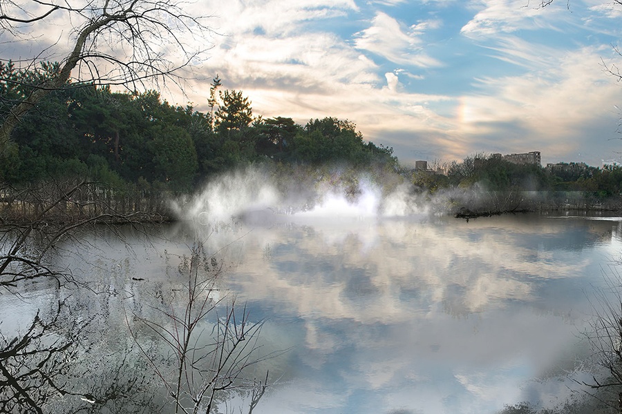
Fog Sculpture rendering in Olmsted Park over an island on Leverett Pond, Brookline, Massachusetts

To celebrate the 20thanniversary of the Emerald Necklace Conservancy Japanese artist Fujiko Nakaya will exhibit five fog works along the historic urban parks that link more than a dozen Boston neighborhoods.

Fujiko Nakaya. Photo courtesy of the Emerald Necklace Conservancy

Nakaya is the daughter of the physicist and science essayist Ukichiro Nakaya, renowned for his work in glaciology and snow crystal photography. Like her father, Ms. Nakaya’s lifelong artistic investigation engages the element of water and instills a sense of wonder in everyday weather phenomena.

Working as part of the legendary group Experiments in Art and Technology (E.A.T.), she first enshrouded the Pepsi Pavilion at the 1970 World Exposition in Osaka in vaporous fog, becoming the first artist to create a sculptural fog environment.

Pepsi Pavilion Osada Japan Photo by Fujiko Nakaya

For the last forty years Nakaya has been partnering with Thomas Mee, a Los Angeles-based engineer.  Mee figured out a system for generating water-based artificial fog. To make it work the installation uses a special fog system that included high-pressure pumps and specifically designed fog nozzles. Several outside factors, like wind conditions, temperature and relative humidity in the environment, determined how intense or thick the fog would be at any given time.

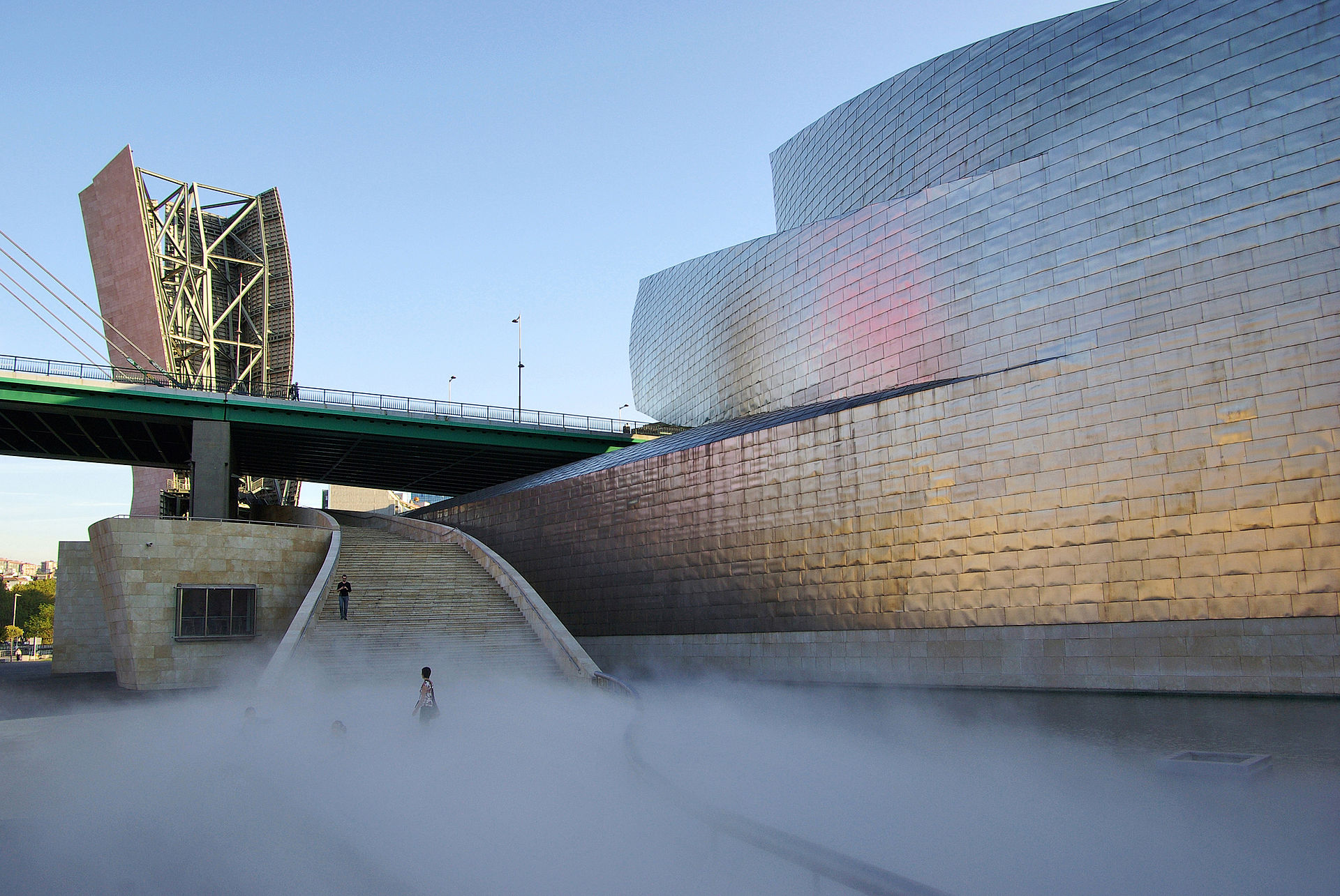
Nakaya has established many other fog installations at galleries worldwide, including the Australian National Gallery, Canberra and the Guggenheim Museum Bilbao, Spain.

Guggenheim Museum Bilbao Photo by Phillip Maiwald

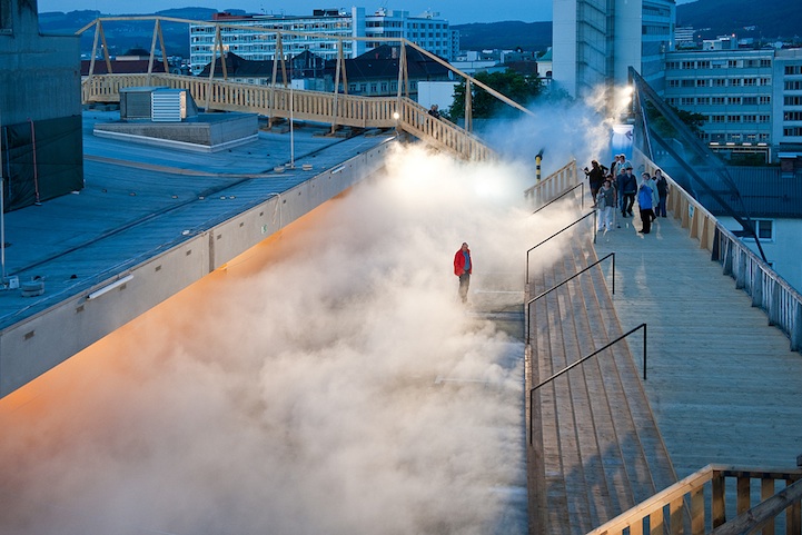
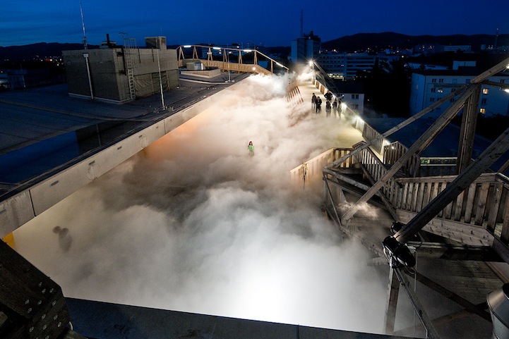
Here are two views of the Cloud Parking by Nakaya located in Linz, Australia – by day and at night:

Daytime photo of Cloud Parking by Fujiko Nakaya

Night View of Cloud Parking

Veil: The Glass House fog installation by Fujiko Nakaya in New Canaan, Connecticut Photo: theglasshouse.org

In Veil – shown above, Nakaya has wrapped the Glass House or Johnson House in a veil of dense mist that comes and goes. For approximately 10 to 15 minutes each hour, the Glass House will appear to vanish, only to return as the fog dissipates. Inside the structure, the sense of being outdoors will be temporarily suspended during the misty spells.

The 85-year-old artist describes her work as a “conversation with nature,”  creating shape-shifting, cloud-like, pure water forms that rhythmically appear and dissipate, inviting visitors to immerse themselves in the art while experiencing the landscape anew.


Hitchhiking to Mars

NASA’s InSight Mars lander is not traveling alone. Two small briefcase size spacecrafts, nicknamed “Wall-E” and “Eva” are hitching a ride as the first CubeSats to visit another planet.

NASA illustration two CubeSats at Mars

A CubeSat is a miniatized satellite for space research made up of multiple cubic units. CubeSats became very popular in the 2000’s for applications such as communications, tracking shipping or performing Earth observation.  Until now, all of them have stayed closed to our home planet.

The Mars twin CubeSats, officially called MarCO-A and B, flew on the same Atlas V rocket that sent InSight into space from Vandenberg Air Force Base in California on May 4, 2018.

Insight lifting off Credit: Ben Smegelsky/NASA

Serving as scouts, the CubeSats will have a front-row seat of the show. Theywill follow InSight on its interplanetary trajectory to Mars and attempt to track the larger spacecraft’s descent and landing on Mars in late November.

Both CubeSats have already phoned home shortly after their release, indicating that their solar panels are providing enough charge in their batteries to deploy their own solar arrays, stabilize themselves, pivot toward the Sun and turn on their radios.

Both MarCos use a compressed gas commonly found in fire extinguishers to push themselves through space, the same way Wall-E did.

Movie still of Wall-E and Eve Credit: Pixar

In addition to charging their own batteries, the twins’ delicate electronics will also need to withstand bursts of radiation on their way to Mars.

If all goes according to plan, InSight will reach its destination in a little less than seven months. If the twins make it they will provide a welcome set of extra eyes as InSight tries to stick its landing on Mars.

This will be a crucial first test of CubeSat technology beyond Earth’s orbit, demonstrating how they could be used to further explore the solar system.

Science Winners

Five gorgeous examples of science photography.

Fluid Mechanics Credit: Stuart Hirth/New Scientist Eureka Prize for Science Photography

The amazing photograph above shows splashes formed from single drops landing in puddles. Captured over several months, they were photographed in darkness using a high-speed flash to preserve their colors and shapes and then brought together in one image.

Liquid Lace Credit: Phred Peterson/New Scientist Eureka Prize for Science Photography.

This winning photograph shows drops of glycerin and water impacting a thin film of ethanol.  The difference in surface tension creates holes in the drop’s surface making it look like lace.

Growth of agaric toadstool mushroom Credit: Phred Petersen/ Royal Photographic Society.

Another image created by Phred Petersen.  This is a time lapse image showing the progress of an agaric toadstool mushroom as it grows.

Phred Petersen is a Senior Lecturer and Coordinator Scientific Photography, School of Media and Communication at RMIT University, a global university of technology and design.

Obelia hydroid Credit: Teresa A. Zgoda

This last photo is a confocal image of a marine organism (obelia hydroid) taken with the 10x objective.  It was a winner from the 2016 International Images for Science competition.

Confocal cross section view of a dandelion showing curved stigma with pollen. Credit: Dr. Robert Markus

Just one more – an honorable mention from 2017 Nikon Small World Competition.


Art of Dark Matter

No one can photograph dark energy itself. But a new camera is looking for the effects of dark energy by gathering data on more than 300 million galaxies whose faint light has been traveling toward Earth for a very long time.

The so called Dark Energy Camera is part of the Dark Energy Survey (DES) and has been fitted to the Victor M. Blanco Telescope located at Cerro Tololo Inter-American Observatory in Chile.  It has been in use since September 2012, with the survey starting in August 2013. This combined optical/near-infrared survey will be used by cosmologists to probe the dynamics of the expansion of the universe and the growth of large-scale structures such as galaxies in their early history.

570 megapixel Dark Energy camera, shown here being tested at Fermilab, will help astronomers uncover the mysteries of dark matter Credit: Image courtesy of Fermilab.

The Blanco telescope in Chile as seen from the air. Credit: NOAO/AURA/NSF

Our universe is not only expanding, but that expansion is speeding up. The faster an object moves away from the Earth, the more its light shifts toward a red color. Measurements of distant galaxies show all of them are red-shifted and moving away from us and from each other. The Dark Energy camera is able to image large swaths of the night sky while accounting for these large red shifts.

The Dark Energy Camera features 62 charged-coupled devices, which record a total of 570 megapixels per snapshot. Credit: Dark Energy Survey Collaborative

The Dark Energy Survey is a five-year effort to map that survey area in unprecedented detail. Scientists will use the data collected to probe the phenomenon of dark energy, the mysterious force that makes up about three-quarters of the universe. The Dark Energy Survey is a collaboration of more than 300 scientists from 25 institutions in six countries. Funding for DES projects are provided by the U.S. Department of Energy Office of Science, the National Science Foundation, and other funding agencies.

The Dark Energy Camera is in its third year of capturing eye-popping images of the cosmos.

Barred spiral galaxy NGC 1365, which lies about 60 million light years from Earth. Credit: Dark Energy Survey Collaboration

Image of NGC 1398 galaxy was taken with the Dark Energy Camera. This galary lives roughly 65 million light years from Earth. It’s slightly larger than our own Milky Way galaxy. Credit: Dark Energy Survey.
Image of NGC 1398 galaxy was taken with the Dark Energy Camera. This galaxy lives roughly 65 million light years from Earth. It’s slightly larger than our own Milky Way galaxy. Credit: Dark Energy Survey.

Comet 2014 Q2 (Lovejoy) on December 27, 2014 happened to be in the field of view of the 570-megapixel Dark Energy Camera © Fermilab
On December 27, 2014 Comet 2014 Q2 (Lovejoy) happened to be in the field of view of the Dark Energy Camera © Fermilab

Ting Li, a Texas A&M astronomy graduate student, developed her own instrument designed to help scientists understand more about the cosmos. Her breakthrough device called the Atmospheric Transmission Monitoring Camera or aTimCam measures subtle changes in the light that is constantly moving through out the atmosphere. The aTimCam has four charge-coupled device cameras equipped with four photographic camera lenses that serve as telescopes individually capture unique images of the wavelengths of light transmitted by a particular star. Li then can track changes in the atmosphere by observing the specific features of each image. The resulting data will be used by scientists as part of the Dark Energy Survey.

Ting Li,testing the Atmospheric Transmission Monitoring Camera installed at Chile’s Cerro Toloto Inter-American Observatory. Credit: © Texas A&M University
Ting Li, testing the Atmospheric Transmission Monitoring Camera installed at Chile’s Cerro Toloto Inter-American Observatory. Credit: © Texas A&M University

Tracking dark matter will show us where our universe has been and hopefully where it will be in the future


The Only Woman You Need to Know

Young Marie Curie Kto8d5W

Ask any stranger to name a female physicist and their response will be Marie Curie.

Manya Sklodovska, as she was called by her family, was born in Warsaw, November 7, 1867 the fourth girl of a moderately successful Polish headmaster who taught his children science, history, music, and poetry. Her mother died of tuberculosis when Marie was ten. After finishing what we would term High School, Marie made a pact with her sister, Bronya.  Since they were too poor for both of them to go to college, simultaneously, Marie would work as a governess (a lowly position) and send money to Bronya, so that Bronya could go to college in Paris.  To the credit of both young women, after Bronya graduated, she reciprocated and supported Marie.  Marie Sklodovska first enrolled in Physics, Mathematics, and Chemistry at the Sorbonne (University of Paris) at the age of 24.  It was 1891.

The search for lab space led to a fateful introduction. Marie mentioned her need for a lab to a Polish physicist who though his colleague, Pierre Curie, might be able to assist her. The meeting between Marie and Pierre would change not only their individual lives but also the course of science. In a civil ceremony in July 1895 they became husband and wife.

Pierre and Marie Curie, 1896  Credit:  Corbis
Pierre and Marie Curie, 1896 Credit: Corbis

Marie Curie (1867-1934) and her husband Pierre continued the work on radioactivity started by Henri Becquerel. In 1898, they discovered two new elements, polonium and radium, which they predicted would display an unprecedented degree of the property that Marie christened “radioactivity”. They name Polonium in honor of Marie’s home country. Marie did most of the work of producing these elements, and to this day her notebooks are still too radioactive to use. She went on to become the first woman to be awarded a doctorate in France. In 1903 they shared the Nobel Prize for Physics with Becquerel.

Pierre and Marie Curie in their lab, 1898  Credit:  Bridgeman Art Library
Pierre and Marie Curie in their lab, 1898 Credit: Bridgeman Art Library

Sadly Pierre Curie was killed by a horse-drawn vehicle in 1906.  Marie, 38-years old, was left with two daughters to raise on her own.

Marie Curie with her daughters Irene and Eve, 1908.  Credit:  Corbis
Marie Curie with her daughters Irene and Eve, 1908.

Marie continued her work after her husband’s death and won a second Nobel Prize for chemistry in 1911.

Marie Curie with her daughter Irene.  Credit:  Art Resource
Marie Curie with her daughter Irene. Credit: Art Resource

Irene (1897-1956) became a nuclear physicist, and worked as her mother’s assistant at the Radium Institute, Paris. In 1935 she shared the Nobel prize for Chemistry with her husband Frederic Joliot, for their work on synthesising new radioactive elements.

Marie was persuaded to embark upon a speaking tour in America and launched a campaign to raise $100,000 from the women of America. Mrs. Herbert Hoover arranged a bequest from the American Association of University Women. Mrs. Edsel Ford sent Marie a car.

The Polish-French Marie Curie became a scientific celebrity whom American woman could see as one of their own.

Madame Curie surrounded by press aboard SS Olympia, May, 1921  Credit:  Image Images
Madame Curie surrounded by press aboard SS Olympic, May, 1921 Credit: Image Images

Marie Curie with her daughters, May 1921  Credit:  Library of Congress/Corbis
Marie Curie with her daughters in New York on the White Star SS Olympic Credit: Library of Congress


With President Harding at the White House on May 1, 1921 after her presented her with a vial of radium.      Credit: Bettmann/Corbis
With President Harding at the White House on May 1, 1921 after he presented her with a vial of radium. Credit: Bettmann/Corbis

Marie died in 1934 due to the exposure of radiation – including carrying test tubes of radium in her pockets during research and her service during World War I in mobile x-ray units (which she created). By the time she died Marie had received no less than 129 awards of national merit.

A 1967 commemorative medal to honor the centennial of Marie Curie’s birth  Credit:  HIP/Art Resource
A 1967 commemorative medal to honor the centennial of Marie Curie’s birth Credit: HIP/Art Resource

Super Resolution Microscopy

E. coli and Shigella sp. bacteria
Fluorescence confocal light micrograph of E. coli and Shigella sp. bacteria (blue) in human cell (green) Credit: Stephanie Schuller/Science Source

Fluorescent micrograph of human insulin producing beta cells  Credit:  Doug Melton/Harvard University.  Photo by B.D. Colen
Fluorescent micrograph of human insulin producing beta cells Credit: Doug Melton/Harvard University. Photo by B.D. Colen

It used to be fact, and even children at school were taught one basic principle of light microscopy: it’s impossible to see things that are smaller than 200 nanometers – that is, a 200 millionth of a millimeter. This was called the diffraction limit. So you might be able to see the shapes of very large bacteria or human cells under your microscope, but you won’t be able to see smaller structures inside of these cells or viruses and certainly no single molecules. This year three new Nobel Laureates in Chemistry – William E. Moerner, Stefan W. Hell and Eric Betzig – have turned it all on its head with the development of super-resolved fluorescence microscopy.

Stefan Hell developed a special method which makes use of two laser beams. One stimulates fluorescent molecules to glow. A second laser beam, though, cancels out any glow caused by the first beam – except for that in a nanometer-sized volume. This way, he yielded images with a resolution much better than conventional confocal light microscopes. He called the new microscopy STED – stimulated emission depletion microscopy.

Working separately Moerner and Betzig, using the same concept of fluorescence and turning individual molecules on and off with laser beam pulses, made it possible to see single molecules only one nanometer big under a microscope.

This image demonstrates the difference in resolution of confocal and STED microscopy. It shows proteins of the nucleus, labeled with fluorescent dyes, imaged with a STED microscope.

Difference between confocual and super resolution view of a human cell.  Credit:  Dr. Dr. Kandasamy Biomedical Microscopy Core University of Georgia.
Difference between confocual and super resolution view of a human cell. Credit: Dr. Dr. Kandasamy Biomedical Microscopy Core University of Georgia.

Since this technology can be used with living cells it will be an invaluable resource when studying HIV or to observe how living cancer cells react to certain cancer drugs. Biologists will even be able to study nerve synapses and the interaction between viruses and cells. This will be a rapidly expanding field in many, many branches of science.

Super-resolution image of a living neuron captured on STED reveals dendritic spines in unprecedented detail.  Photo by Stefen Hell from the Max Planck Institute on Biophysical Chemistry.
Super-resolution image of a living neuron captured on STED reveals dendritic spines in unprecedented detail. Photo by Stefen Hell from the Max Planck Institute on Biophysical Chemistry.

Protein network in a mammalian cell captured with STED Photo from Max Planck Institute of Biophysical Chemistry

To watch a video on the STED microscope produced by the Max Planck Institute click below.

MPI Images of Science

The Max Planck Society is Germany’s most successful research organization. Since its establishment in 1948, no fewer than 17 Nobel laureates have emerged from the ranks of its scientists. The currently 82 Max Planck Institutes conduct basic research in the service of the general public in the natural sciences, life sciences, social sciences, and the humanities. They themselves define their research subjects and are given the best working conditions, as well as free reign in selecting their staff.  Recently the Max Planck Society has been establishing a Partner Institute in Shanghai and the first Max Planck Insitute in North America financed by the State of Florida.

Here are some colorful images of science from their online gallery.

Scanning electron microscope image of white blood cells (shown here in red) encircling tuberculosis bacteria (shown in yellow).  © Max Planck Institute for Infection Biology / Volker Brinkmann
Scanning electron microscope image of white blood cells (shown here in red) encircling tuberculosis bacteria (shown in yellow).
© Max Planck Institute for Infection Biology / Volker Brinkmann

Electrons at point of contact in a two-dimensional semiconductor.  The lower half shows ideal transverse; the up half of image show how strongly a weak disorder in the material can effect the electrons’ path.   © Max Planck Institute for Dynamics and Self-Organization / Ragnar Fleischmann
Electrons at point of contact in a two-dimensional semiconductor. The lower half shows ideal transverse; the up half of image show how strongly a weak disorder in the material can effect the electrons’ path.
© Max Planck Institute for Dynamics and Self-Organization / Ragnar Fleischmann

Light micrograph with polarized light showing the microstructure of cast iron Credit: © Max-Planck-Institut für Eisenforschung GmbH, Düsseldorf / Angelika Bobrowski
Light micrograph with polarized light showing the microstructure of cast iron
Credit: © Max-Planck-Institut für Eisenforschung GmbH, Düsseldorf / Angelika Bobrowski

Confocal laser-scanning microscope image of proteins interacting with lipid membrane.  Credit: © Max Planck Institute of Biochemistry, Martinsried / Katja Zieske, Petra Schwille
Confocal laser-scanning microscope image of proteins interacting with lipid membrane. Credit: © Max Planck Institute of Biochemistry, Martinsried / Katja Zieske, Petra Schwille

Scanning electron microscope image of powdery mildew on leaf © Max Planck Institute for Developmental Biology / Jürgen Berger, Marco Todesco
Scanning electron microscope image of powdery mildew on leaf
© Max Planck Institute for Developmental Biology / Jürgen Berger, Marco Todesco