Feminist History of Illustrating Plants

Most people are unaware of several talented women who worked in the field of botany as early as the 18thcentury. 

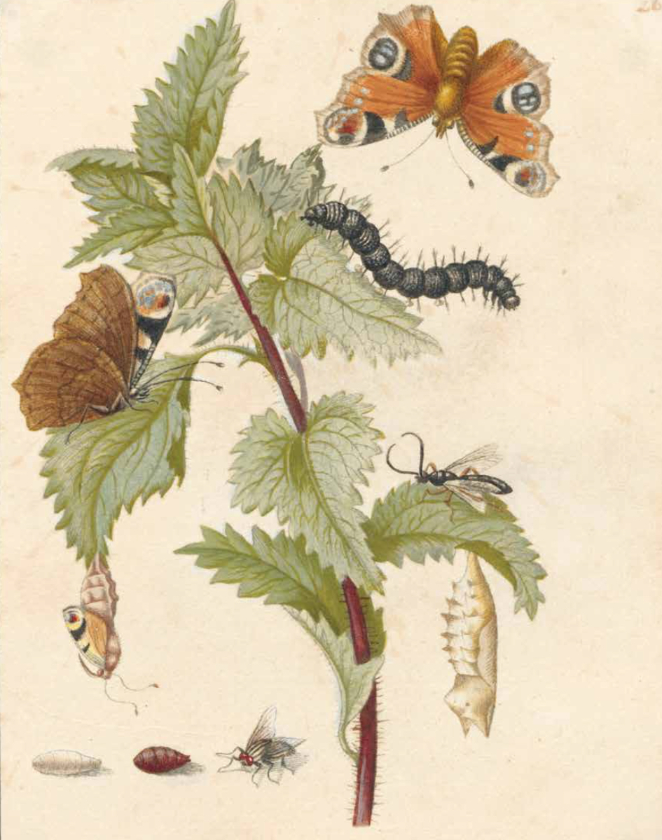
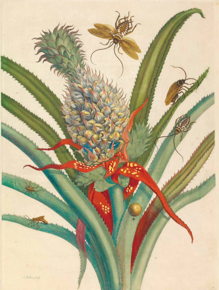
Consider Maria Sibylla Merian (1647-1717) Maria was an extremely enterprising and independent German woman.  In 1699, along with her daughter, she travelled to Surinam to carry out research into the reproduction and development of insects. She is now regarded as both a highly gifted artist and an exceptional empirical scientist, one of the first to demolish the prevailing notion of the spontaneous generation of insects from mud.

Portrait of Maria Sibylla Merian

Since women were not allowed to sell paintings in oils in many German cities, Maria became skilled at watercolor and gouache. She was the first to portray caterpillars and butterflies with the plants that nourished them.  Maria’a book, Metamorphosis insectorum Surinamensium,was published in 1705 in both Latin and Dutch with colored engravings.  Marian paid the production costs her self and acted as the publisher.  

Maria’a book, Metamorphosis insectorum Surinamensium,was published in 1705 in both Latin and Dutch with colored engravings.  Marian paid the production costs her self and acted as the publisher.  Two folio editions of 254 aquarelles by Marian were taken to Saint Petersburg for Peter the Great’s personal physician.

Ananas mit Kakerlake (Pineapple with cockroach) by Merian (c. between 1701 and 1705) Hand coloured copper engraving


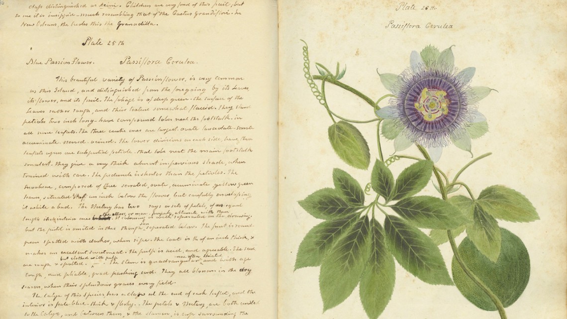
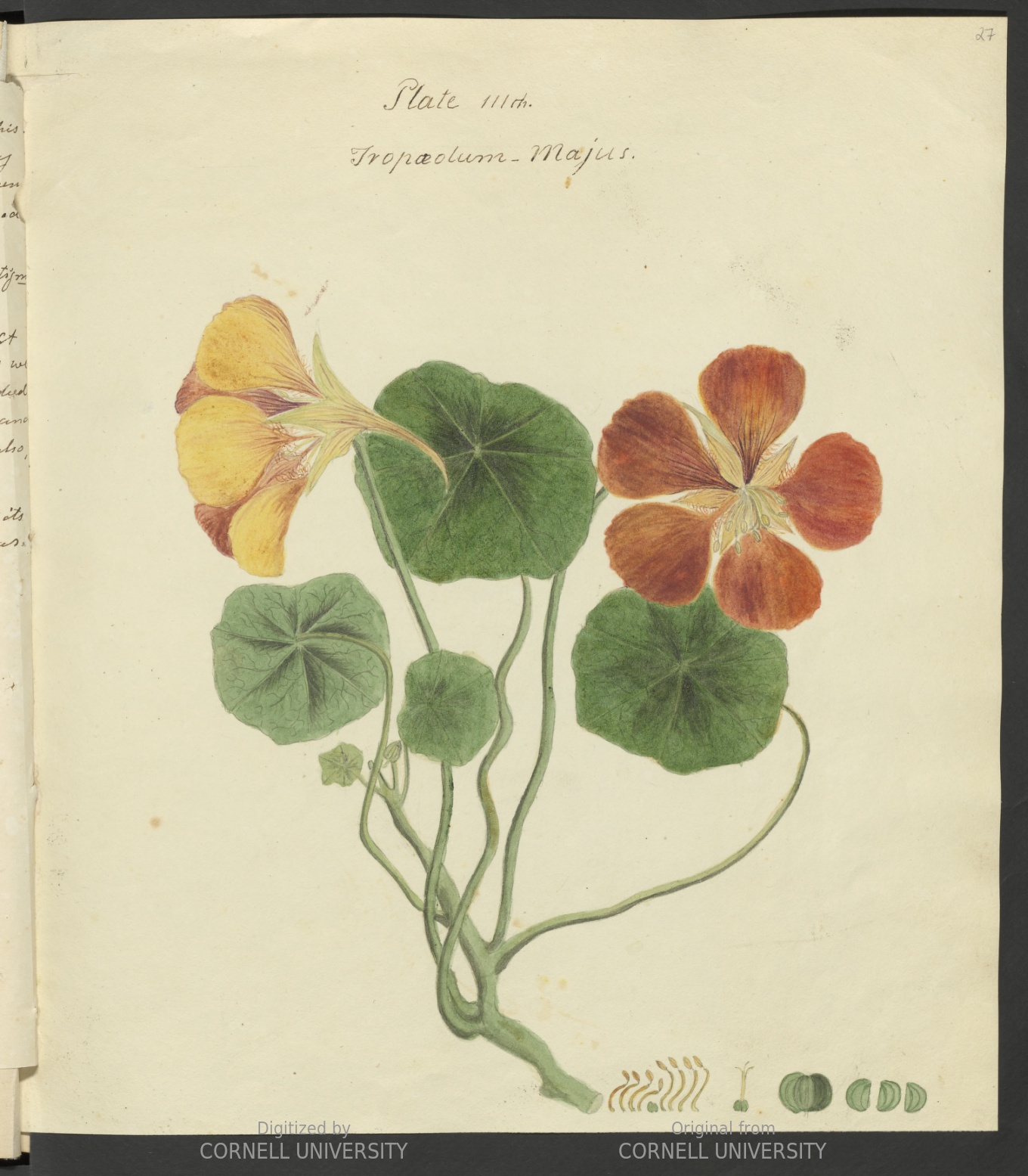
Anne Kingsbury Wollstonecraft (October 29, 1791 – May 16, 1828) was an American botanist who devoted herself to creating richly detailed illustrations and descriptions of the botanical specimens she found on Cuba. Her work culminating in a remarkable three-volume manuscript entitled, Specimens of the Plants & Fruit of the Island of Cuba. This book was never published and went missing for 190 years. It was recently discovered at Cornell Library’s division of rare manuscripts.  The book includes 121 watercolor plants with detailed notes.

Page from Specimens of the Plants & Fruits of the Island of Cuba by Anne Kingsbury Wollstonecraft.  Credit:  Cornell University.
Anne Kingsbury Wollstonecraft   Credit: Cornell Division of Rare Manuscripts

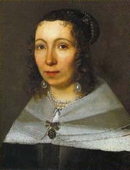
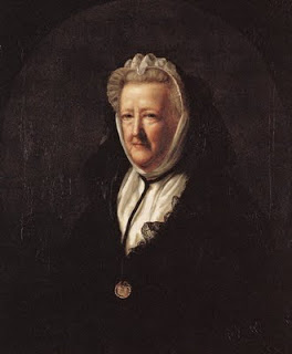
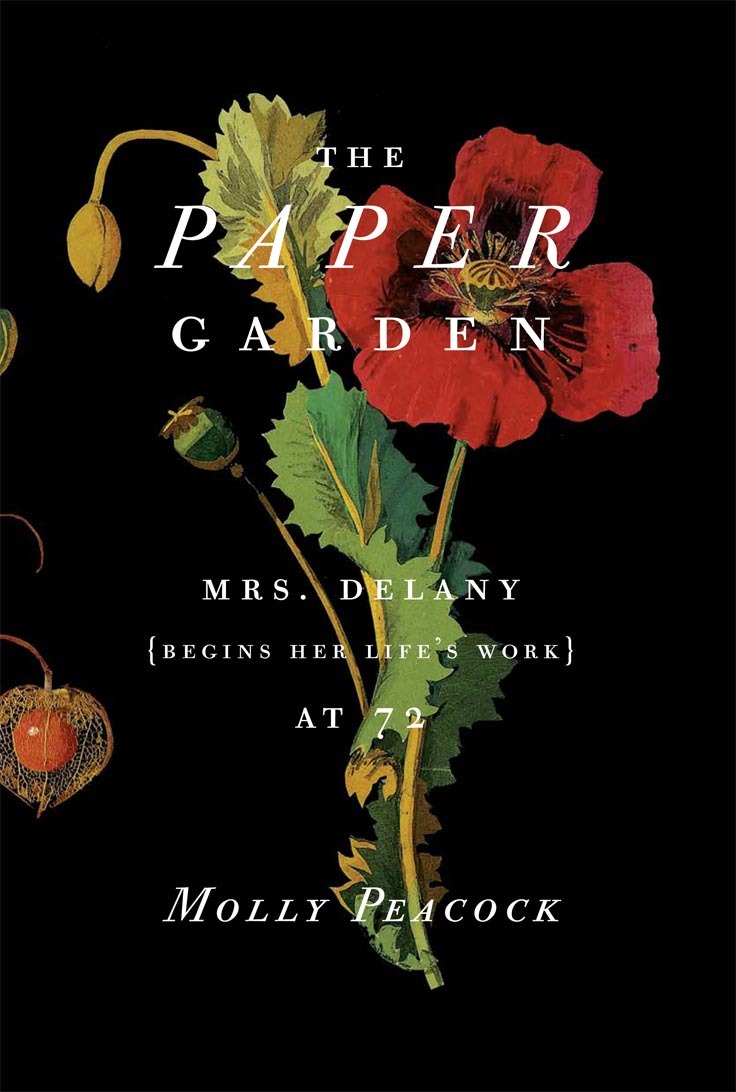
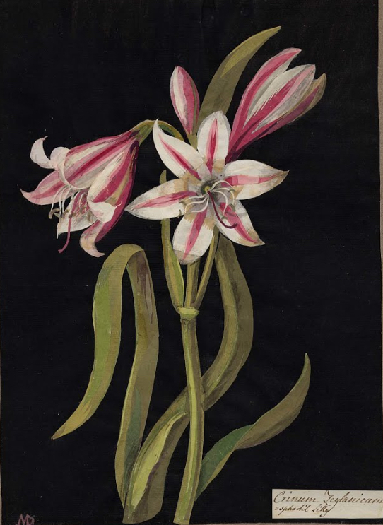
Just as remarkable was Mary Delany (May 14, 1700- April 15, 1788), an English woman whose collection of intricate paper collages of plant life are now in the British Museum.  Mary Delany created dramatic and precise collages, made from colored paper, much of which she had dyed herself. The works were then mounted on black backgrounds. Describing her method in a letter to her niece dated October 4th, 1772, she wrote: “I have invented a new way of imitating flowers”. She was then 72. In ten years times Mary Delaney completed nearly 1,000 cut-paper botanicals so accurate that botanists still refer to them – each one so energetically dramatic that it seems to leap out from the dark as on to a lit stage.

Portrait of Mary Delany by John Opie, 1782 
Asphodi Lilly paper collage by Mary Delany     Photo: © Trustees of the British Museum

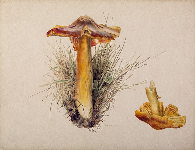
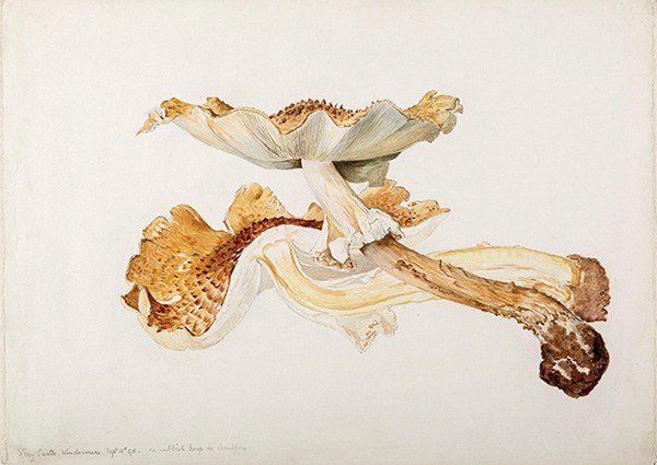
Beatrix Potter (July 28, 1866 –December 22, 1943), famous for The Tale of Peter Rabbitand other children’s books, has been underappreciated for her contribution to science and natural history.  In her early twenties, Beatrix developed a keen interest in mycology and began producing incredibly beautiful drawings of fungi.  She taught herself the proper technique for accurate botanical illustration.  

When she wanted to present her scientific work to London’s Linnean Society she needed to have her uncle do the presentation because women were barred from membership.  The paper never got peer-review and was dismissed as not worthy of consideration. A century later the Linnean Society apologized for its historic sexism. 

Beatrix Potter’s drawing of Hygrophorus puniceus Credit:   Armitt Museum and Library
Beatrix Potter’s drawing of Lepiota friesii   Credit:  Armitt Museum and Library

The female amateur botanists and naturalists of earlier eras didn’t just reproduce knowledge. They took what they learned and used the traditionally feminine skills they already had—along with their keen powers of observation—to create something better, and new.

Father of Crop Art

Stan Herd Photo courtesy of Jon Blumb

A native of Protection, Kansas, Stan Herd nourished a love of art as a young man and was awarded an art scholarship to Wichita State University in 1969.

After working in New York for a number of years he returned to his heritage in rural Kansas and began developing a large quantity of artwork including paintings and large murals with mostly western imagery. A few years into his career as an established western artist, Herd had a thought.

“I was on a flight back from Dodge City in 1976 after painting a mural on a bank when I was looking down at a field and saw a tractor traversing a field corner to corner when the idea came to me to create a design on the ground,” he explained. “

Herd’s first Earthwork was of Chief Satanta, a Kiowa leader.  It was several years in the making before it was unveiled in 1981. The 160-acre portrait would become the first of many Earthworks.

Chief Satanta Earthwork and photo by Stan Herd

To date Herd has created about 40 commissioned commercial pieces and 40 art pieces of his own.  The commercial works afford him the freedom to do the work that means the most to him.

He has used combines, tractors, Roto tillers, drills, and many hand-held tools combining new and existing vegetation to carve out an image.  His work sometimes includes mulch, rocks and stones as well. Herd said he usually has a dozen people assist on an average Earthwork project, sometimes family, friends, locals, students and agriculturists who know the area and its crops.

Stan Herd Amelia Earhart as seen from the air. Photographer: TALIS BERGMANIS Credit: THE STAR Keyword: ART

An example of Stan Herd’s commercial work for Shock Top Belgian Ale. This one is in Austin Texas. It is made with two ingredients in the ale: wheat and oranges.

In 2018 Herd received an email from a tobacco executive in China inviting him to visit Yunnan province and see if he wanted to participate in the construction of a public park. After a bit of consideration and recovering from the surprise of the invitation, Herd jumped at the chance. This would be a four-acre earthwork on a hillside in the center of the 800-acre Taiping Lake Park.

Thus began an epic undertaking that found Herd traveling to China 15 times in 15 months. He insisted on creating the design outline without a GPS – laying every single line by himself.”  The work was interrupted by several rainy seasons.

Herd used more than 15,000 bricks chipped out of locally quarried rock. These were placed to create beds for flowering plants and medicinals native to the region. Together the stone and vegetation formed the patterns of the subject’s face and clothing.

Construction Young Woman of China in Yunnan Stan Herd

Herd’s comment about the finished art: “It beautifully represents the elegance of the (women) of China. … And this artwork also (embodies) communication in culture and art areas between China and America.”

Stan Herd LEAD woman in china

Show above is Stan Herd’s Young Woman of China.

Stan Herd’s most recent project is a 1.2-acre recreation of Van Gogh’s famous artwork, Olive Trees, which he “planted” in Minneapolis. The piece was commissioned by the Minneapolis Institute of Art and involved weeks of mowing, digging, planting, and earthscaping to create the piece viewable from the air near the Minneapolis airport. The field location was specifically chosen so that flight passengers can easily see the land art.

Olive Trees by Stan Herd. Van Gogh’s signature dazzling painted sky was recreated using a field of oats mowed in concentric circles.

Olive Trees by Stan Herd as seen from a plane.

Like many of his artworks, Herd’s rendition of Van Gogh’s painting will disappear over time as the crops grow out and the elements wear down the design.

Young Woman of China might not be big enough to be seen from space, however, it does have one advantage over the Great Wall of China.  As Herd explains: “It’s so deep and embedded in the ground, and so massively created on that hillside, it will be here hundreds of years from now.”

Stan Herd has certainly created a niche in the art world.  Dan Rather reporting on CBS News called him the Father of Crop Art.

Pollen Grains by Micronaut

Best known as a scientific photographer, Martin Oeggerli takes close-up images of microscopic creatures and structures that have been featured in scientific publications and art galleries worldwide.

Most of Oeggerli’s best images are taken with a scanning electron microscope (SEM). SEM is always black and white because it uses electrons instead of photons to view the specimen, and only light carries color information.  As an artist, Oeggeril wants the images to be attractive. He tries to highlight morphologically different structures to make them more visible so that the viewer can recognize complexity. And he goes to great lengths to reproduce the original color.

Oeggerli, otherwise known as Micronaut, has collected and explored pollen grains.  Here are a few examples from his gallery:

Water cabbage (Pistia sp.) is an invasive plant. Despite a tiny flower, the pollen is a medium sized elliptic monad covered with equatorial crests, which are rarely found features in the pollen universe. (c) Micronaut

Forget Me Not pollen (c) Micronaut

Pollinium of an orchid, consting of many hundred single pollen grains Credit: (c) Micronaut

Purple willow pollen (c) Micronaut

Pink thrift pollen (c) Micronaut

Pollen on pistil of a geranium (c) Micronaut

One image might take 20-60 hours to create depending on how much detail and how many structures are in the picture.  Because Micronaut goes to great lengths to reproduce the original color each one is a work of art.

Magic of Photomicrography

Polish photographer, Marek Miś, has developed a technique for photographing living and inanimate microorganisms, plant tissues and micro crystals directly through the eyepiece of a microscope. You may have seen his awarding winning photographs in either the Nikon Small World Competition or the Olympus BioScapes Competition.  Here are a few of my favorites:

Crustacean (Nauplius) Polarized light micrograph by Marek Mis
Crustacean (Nauplius) Polarized Light by Marek Mis

Abstract of the drug Rywanol by Marek Mis

Longitudinal stem cross-section of nettle polarized light micrograph by Marek Mis
Longitudinal stem cross-section of nettle polarized light by Marek Mis

Rotifer created using Rheinberg illumination by Marek Mis

Polarized light micrograph showing a sectioned stem of a young apple tree plant Marek Mis
Polarized light micrograph showing a section stem apple tree plant Marek Mis

Desmid (Micrasterias) by Marek Mis

Marek’s microphotographs are made using various lighting techniques – bright field, dark field, phase contrast, Rheinberg illumination, polarized light, oblique illumination and mixed techniques.  To see more of Marek Mis’s work go to this URL: http://www.mismicrophoto.com/plants.php

Confocal microphoto of a barnacle appendage by Igor Siwanowicz

German photographer, Dr. Igor Siwanowicz who works at the Max Planck Institute of Neurobiology specializing in invertebrate photography, has succeeded in showing the world there is beauty of form in the insect world.

Confocal microphoto showing detail of a foot of a giant diving beetle by Igor Siwanowicz

You can see a more complete portfolio of Igor Siwanowicz’s work here:

http://photo.net/photodb/member-photos?user_id=1783374

There is a indeed a whole magical world of images to be seen and created through a microscope.

Seeing Flowers


Macro photo of daffodils by Robert Llewellyn

Here’s a special book to add to your library… Seeing Flowers: Discover the Hidden Life of Flowers, Photography, by Robert Llewellyn, written by Teri Dunn Chace.  The book features over three hundred extraordinary photographs of flowers, organized by family, from the Amaryllis family through the Viola family.  These exquisite macro photographs of flowers are completely addictive.

Llewellyn uses “image stacking”, where multiple shots of a subject are taken at varying focus points and then melded together in a computer application. He shoots as many as 100 images for a single photograph, each image focusing on planes separated by perhaps only a centimeter. Llewellyn’s camera is attached to a computer-controlled motor-driven mount on a vertical axis above his subject. The results are luminous, delicate portraits with every last hair and pollen grain in focus.

Macro photo by Robert Llewellyn

Macro photo by Robert Llewellyn


Macro photo by Robert Llewellyn

Macro photo by Robert Llewellyn

Macro photo by Robert Llewellyn

To see more of Robert Llewellyn’s photography go to

http://www.robertllewellyn.com

MPI Images of Science

The Max Planck Society is Germany’s most successful research organization. Since its establishment in 1948, no fewer than 17 Nobel laureates have emerged from the ranks of its scientists. The currently 82 Max Planck Institutes conduct basic research in the service of the general public in the natural sciences, life sciences, social sciences, and the humanities. They themselves define their research subjects and are given the best working conditions, as well as free reign in selecting their staff.  Recently the Max Planck Society has been establishing a Partner Institute in Shanghai and the first Max Planck Insitute in North America financed by the State of Florida.

Here are some colorful images of science from their online gallery.

Scanning electron microscope image of white blood cells (shown here in red) encircling tuberculosis bacteria (shown in yellow).  © Max Planck Institute for Infection Biology / Volker Brinkmann
Scanning electron microscope image of white blood cells (shown here in red) encircling tuberculosis bacteria (shown in yellow).
© Max Planck Institute for Infection Biology / Volker Brinkmann

Electrons at point of contact in a two-dimensional semiconductor.  The lower half shows ideal transverse; the up half of image show how strongly a weak disorder in the material can effect the electrons’ path.   © Max Planck Institute for Dynamics and Self-Organization / Ragnar Fleischmann
Electrons at point of contact in a two-dimensional semiconductor. The lower half shows ideal transverse; the up half of image show how strongly a weak disorder in the material can effect the electrons’ path.
© Max Planck Institute for Dynamics and Self-Organization / Ragnar Fleischmann

Light micrograph with polarized light showing the microstructure of cast iron Credit: © Max-Planck-Institut für Eisenforschung GmbH, Düsseldorf / Angelika Bobrowski
Light micrograph with polarized light showing the microstructure of cast iron
Credit: © Max-Planck-Institut für Eisenforschung GmbH, Düsseldorf / Angelika Bobrowski

Confocal laser-scanning microscope image of proteins interacting with lipid membrane.  Credit: © Max Planck Institute of Biochemistry, Martinsried / Katja Zieske, Petra Schwille
Confocal laser-scanning microscope image of proteins interacting with lipid membrane. Credit: © Max Planck Institute of Biochemistry, Martinsried / Katja Zieske, Petra Schwille

Scanning electron microscope image of powdery mildew on leaf © Max Planck Institute for Developmental Biology / Jürgen Berger, Marco Todesco
Scanning electron microscope image of powdery mildew on leaf
© Max Planck Institute for Developmental Biology / Jürgen Berger, Marco Todesco

What’s the DOE Up to?

Reaction of minerals with CO2   Credit:  Pacific National Laboratory/US Department of Energy
Reaction of minerals with CO2 Credit: Pacific Northwest National Laboratory/US Department of Energy

Pacific Northwest National Laboratories, funded by the U.S .Department of Energy’s Office of Basic Energy Sciences, is studying how minerals react to carbon dioxide gas at low temperatures.  .  This image was captures with a helium ion microscope and shows two minerals (forsterite in gray and blue and dypingite in orange and yellow) after reacting with fluid-like supercritical CO2 for 43 days at 50 degrees Celsius.   Why?  To inform efforts to capture and store CO2 and other greenhouse gases deep underground.

Penicillium fungus on plant roots.  Credit:  PNNL/DOE
Penicillium fungus on plant roots. Credit: PNNL/DOE

National Science Foundation and U.S. Department of Energy are funding a study of biogeochemistry of plant-microbe-soil interactions.   The work  is expanding our understanding of how to provide nutrients to plants by mineral dissolution assisted by microbes.

Nanofibers captured with a helium ion microscope.  Credit: PNNL/DOE
Nanofibers captured with a helium ion microscope. Credit: PNNL/DOE

Nanofibers are an exciting new frontier of science.  The fibers shown in the photo above, created by team members from Pacific Northwest National Laboratory, are helping to advance the design of novel metal alloy anodes.  The anodes are expected to improve the effectiveness of sodium-ion batteries and to lead to low-cost energy storage solutions for the nation.  The research is funded by the U.S. Department of Energy’s Basic Energy Science program.

Zinc oxide nanostructures.  Credit:  PNNL/DOE
Zinc oxide nanostructures. Credit: PNNL/DOE

Zinc oxide is a unique and versatile semiconductor material that’s attracting attention due to its potential applications in a wide range of technologically important fields, including electronics, spintronics and radiation detection.  Nanostructures of zinc oxide offer promise for miniaturizing electronic devices.  The image was captured with a helium ion microscope.  The project is funded by the DOE’s Office of Biological and Environmental Research with additional research partners at Oak Ridge National Laboratory and the University of Tennessee, Knoxville.

Topiaries Far Beyond the Skill Set of Edward Scissorhands

The Montréal Botanical Garden has been offering its millions of visitors an unforgettable experience for over 80 years.  This summer The International Mosaiculture event is back after a ten-year absence on view at the Botanical Garden from June 22 to September 29, 2013.

This incredible display of two and three-dimensional constructions, cultivated by 200 international horticultural artists originating from more than 20 countries, makes it the largest eco-responsible event to come to Quebec.

Mosaiculture is a multifaceted discipline, drawing on a range of craftsmanship and knowledge — sculpture for the framework, painting for chroma, and ecology, for the understanding of the maintenance of the floral medium. This year’s event challenged the artists around the theme ‘land of hope’ as it reflects their own culture, drawing influences from icons of peace and promise for a environmentally sound world. Collectively, over three million colorful flowers and plants were used in the environmental designs, creating a colossal body of living vibrant art.

1683384-inline-s-6-edward-scissor-hands-type-botanical-garden-designs

http://vimeo.com/70414850

247871_306024_detail_eplv

The man who planted trees.  Photo from Mosaecultures-Internationales-de-Montreal 2013
The man who planted trees. Photo from Mosaecultures-Internationales-de-Montreal 2013

Montreal plant

plant-sculptures-mosaicultures-internationales-de-montreal-3

166699_detail_eplv

Biomedical Image Awards

Each year Wellcome Images puts its researchers to the task of finding the most amazing biomedical photography.  My favorites are always micrographs.  I’ve paired a few award winning images with the source of each image to better explain the magnification:

SEM lavender leaf.  Credit:  Wellcome Images
SEM lavender leaf. Credit: Wellcome Images

Lavender leaves.  Credit:  Mill Creek Gardens LLC
Lavender leaves. Credit: Mill Creek Gardens LLC

In the top photo a false-coloured scanning electron micrograph (SEM) shows a lavender leaf  imaged at 200 microns (approximately 0.0079  inches).  Lavender yields an essential oil with sweet overtones, which can be used in balms, salves, perfumes, cosmetics and topical applications. It is also used to aid sleep, to relax and to alleviate anxiety. The surface of the leaf is covered with fine hair-like outgrowths made from specialized epidermal cells called non-glandular trichomes, which protect the plant against pests and reduce evaporation from the leaf. Glandular trichomes are also present, containing the oil produced by the plant.

African clawed frog eggs Credit:  Vincent Pasque/Wellcome Images
African clawed frog eggs Credit: Vincent Pasque/Wellcome Images

African clawed frog
African clawed frog

African clawed frog oocytes (immature egg cells) are shown in the top photo.  Each oocyte is surrounded by thousands of follicle cells highlighted by staining DNA blue. Blood vessels, which provide oxygen to the immature egg cells and follicle cells, are shown in red. The ovary of each adult female African clawed frog contains up to 20,000 oocytes.  At approximately 1.2 mm in diameter they are much larger than the eggs of many other species.

Micrograph of aspirin crystals.  Credit:  M.I. Walker/Wellcome Images
Micrograph of aspirin crystals. Credit: M.I. Walker/Wellcome Images

Single aspirin
Single aspirin

In the top image, crystals of aspirin, that common drug used to relieve minor aches and pains, look more like psychedelic visuals caused by a totally different kind of drug.

Bird of paradise seed. Credit:  Annie Cavanagh/Wellcome Images
Bird of paradise seed. Credit: Annie Cavanagh/Wellcome Images

Bird of Paradise flower

The top photograph of a seed was taken using an electron microscope.  This strange looking seed is from a bird of paradise plant, which is native to South Africa and has a distinctive orange and blue flower, which resembles an exotic bird.

SEM of microparticle drug.  Credit:  Annie Cavanagh/Wellcome Images
SEM of microparticle drug. Credit: Annie Cavanagh/Wellcome Images

Co-polymer capsules
Co-polymer capsules

The orange and blue balls shown in the top photo represent electron microscope images of co-polymers. These are used for a drug delivery system known as particle in particle. The inner particle, shown in orange, is loaded with the drug prednisolone, used to treat inflammatory bowel disease. The outer particle, in blue, is a co-polymer which encapsulates the inner particle. Polymers can be used to coat a drug to prevent it being released in the stomach, or to produce a slow release of drug.